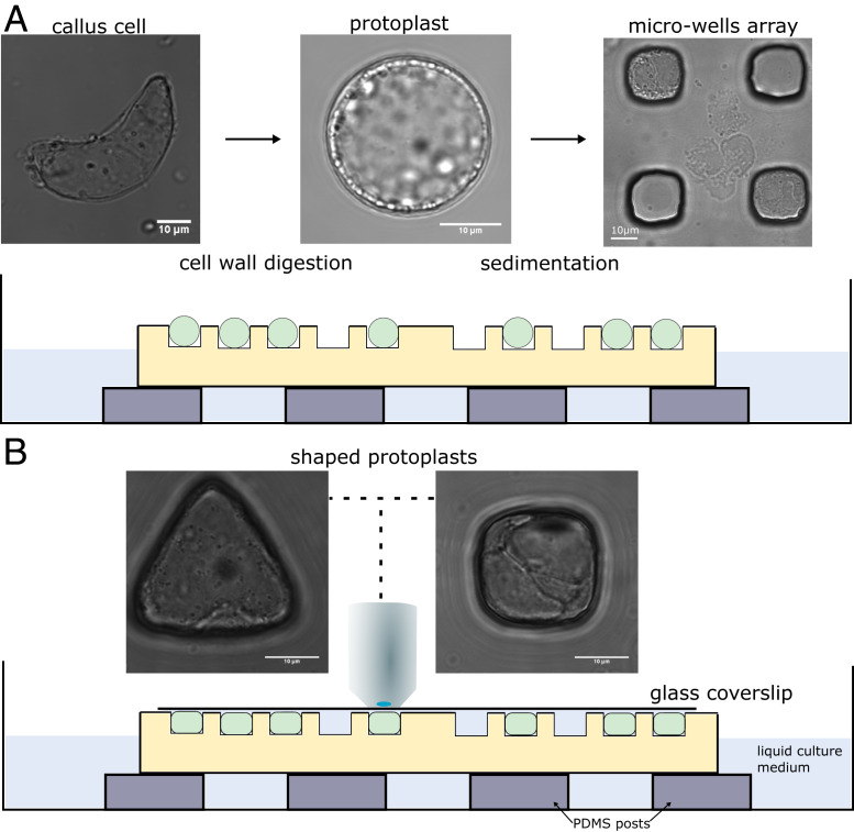
Fig. 1.
Description of the experimental setup used for the confinement of protoplasts in different geometries. (A) The cell walls of Arabidopsis thaliana root-derived callus cells were digested to make spherical protoplasts. Protoplasts were then plated on top of the microwell array. (Left) Bright-field picture of an isolated cell from callus with undefined geometry. (Middle) Bright-field picture of a freshly isolated protoplast with spherical geometry. (Right) Bright-field picture of protoplasts in square microwells after sedimentation. (Bottom) Illustration of microfluidic setup with cells loaded in wells. (B) Coverslips were carefully placed on top of the microwells, and the samples were imaged using an upright microscope. Bright-field pictures of protoplasts being shaped in microwells of various geometries (63× objective). All size standards are 10 µm.

