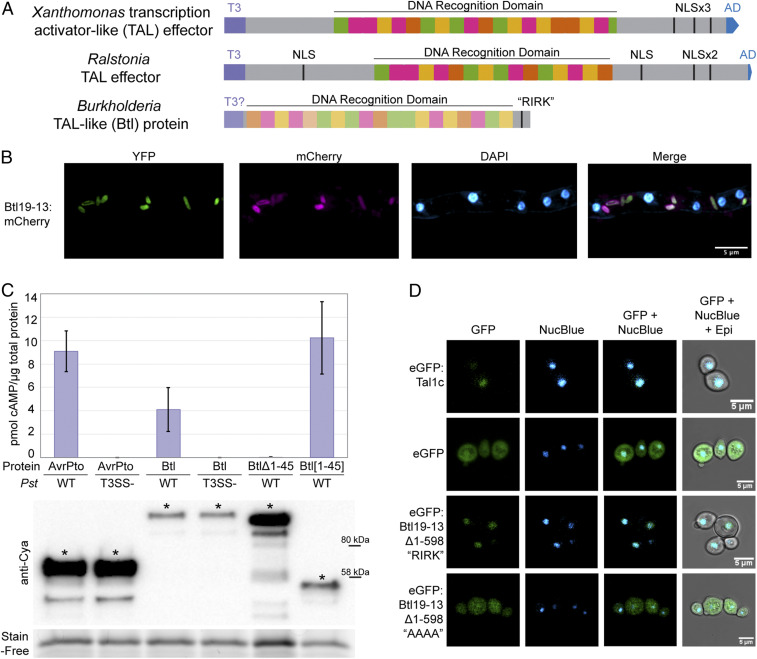
Fig. 1.
Structure, expression, T3 secretion, and nuclear localization of Btl19-13. (A) Domain structure of a Xanthomonas TAL effector, a Ralstonia TAL effector, and a Btl protein. T3, type III secretion signal; T3?, putative type III secretion signal; NLS, nuclear localization signal; “RIRK,” aa sequence of a putative NLS in Btl19-13; AD, activation domain. (B) Confocal microscopy of Mycetohabitans sp. B13 cells constitutively expressing EYFP and expressing Btl19-13:mCherry under the native btl19-13 promoter, within a R. microsporus hypha. DAPI staining of the nuclei is included for reference. (C) Quantification of cAMP by ELISA in Nicotiana benthamiana leaf punches 6 h after infiltration with Pseudomonas syringae pv. tomato DC3000 (Pst) expressing the following: AvrPto:Cya (61 kDa), Btl19-13:Cya (118.6 kDa), Btl19-13:Cya missing the first 45 aa (Btl19-13Δ1-45, 110 kDa), and the first 45 aa of Btl19-13 fused to CyaA (Btl19-13[1-45], 51 kDa). T3SS designates an hrpQ-U mutant incapable of type III secretion (28). Each bar shows the results from three biological replicates with two technical replicates each. Error bars denote SD. The experiment was repeated twice with similar results. Below, a Western blot of leaf homogenates probed with anti-Cya, shown with a Stain-Free loading control (Bio-Rad). Asterisks (*) indicate bands corresponding to the expected size. (D) Confocal microscopy of Saccharomyces cerevisiae cells expressing the indicated proteins and stained with NucBlue to locate nuclei. Protein expression was induced with galactose 24 h prior to imaging. “AAAA” indicates an alanine substitution of the “RIRK” motif in Btl19-13 (residues 692–695).

