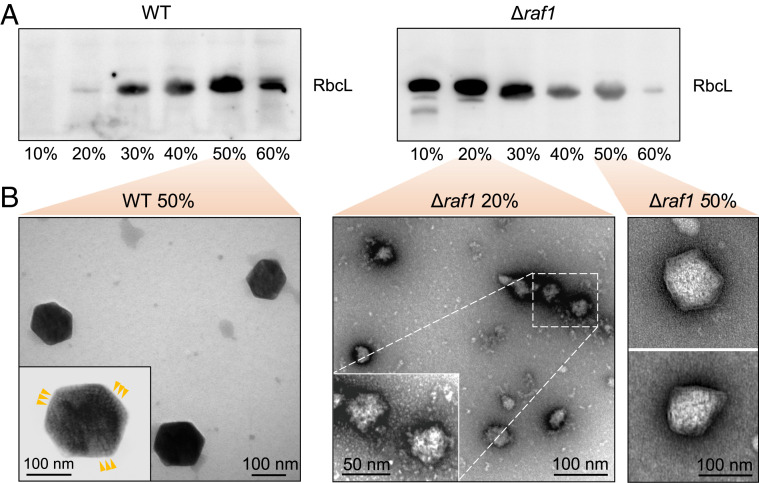
Fig. 5.
Characterization of the carboxysome assembly assemblies. (A) Relative Rubisco amount in each sucrose gradient fraction achieved during carboxysome isolation from the WT (Left) and Δraf1 mutant (Right), as revealed by SDS-PAGE and immunoblot analysis using an anti-RbcL antibody. (B) Negative-staining TEM images of intact carboxysomes in the 50% fraction from WT (Left) and assembly intermediates in the 20% and carboxysome-like structures in the 50% fractions from the Δraf1 cells (Right). (Insets) Zoom-in views representing the structures and protein organization of an intact carboxysome from the WT and the assembly intermediates from the Δraf1 mutant. Arrows indicate the ordered Rubisco arrays observed in the WT carboxysomes.

