
Figure 4. Validation of MAP4K Protein Interaction Network.
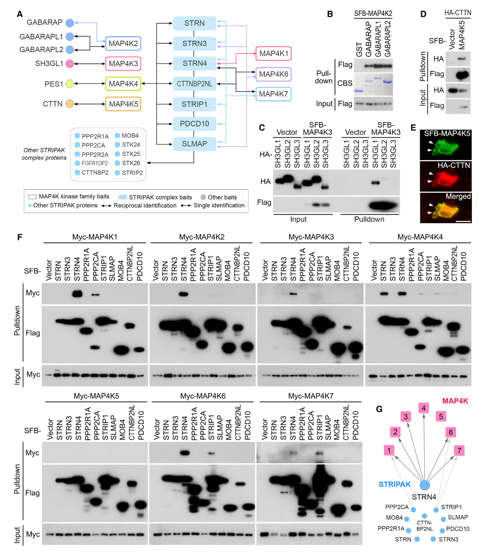
(A) A summary of reciprocal TAP-MS analyses for the selected MAP4K-HCIPs.
(B) Validation of the interaction between MAP4K2 and GABARAP family proteins. The bacterially purified GST-GABARAP family proteins were subjected to the pull-down assay. CBS, Coomassie blue staining.
(C) Validation of the interaction between MAP4K3 and SH3GL1. HEK293T cells were transfected with the indicated constructs and subjected to the pull-down assay.
(D and E) Validation of the interaction between MAP4K5 and CTTN. HEK293T cells were transfected with the indicated constructs and subjected to the pull-down assay (D). The co-localization between MAP4K5 and CTTN in lamellipodia was indicated by arrows (E). Scale bar, 20 μm.
(F and G) Validation of the interaction between MAP4K family kinases and the STRIPAK complex components. HEK293T cells were transfected with the indicated constructs and subjected to the pull-down assay (F). The pull-down experiment results were summarized (G).