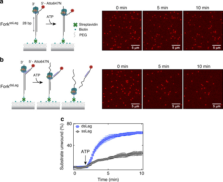
Fig. 4. Single-molecule analysis of CMG-catalyzed fork unwinding.
a, b Visualizing CMG-driven unwinding of individual Atto647N-labeled surface-immobilized fork substrates with TIRF microscopy. Unwinding of 28-bp duplex region by CMG leads to dissociation of the fluorescent strand from the surface. Representative fields of view are shown at three time points on ForkssLag a and ForkdsLag b following ATP addition (n = 3 independent experiments). c Percentage of molecules unwound as a function of time for ForkssLag (gray, N = 1215 molecules from n = 3 independent experiments) and ForkdsLag (blue, N = 1611 molecules analyzed). Data represent mean ± SD from three independent experiments for each substrate. Source data are provided as a Source Data file.

