Figure 4.
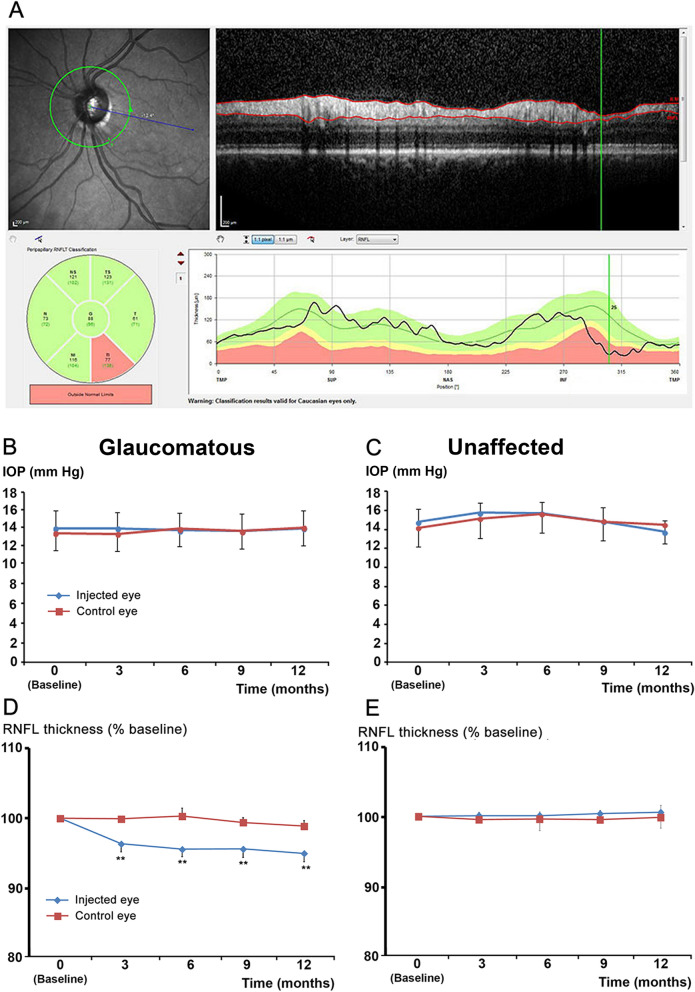
Treatment with VEGF trap reduces RNFL thickness in glaucomatous patients. (A) Circular peripapillary OCT scan analysis: An abnormal left eye with its corresponding fundus image (upper left). Red lines in the OCT B-Scan (upper right) indicate the inner and outer borders of the RNFL found by the algorithm. RNFL thicknesses (lower right) plotted over the thickness values measured in healthy subjects of the same age. Mean thickness of the RNFL in the six sectors (lower left). This scan shows a significant decrease (red segment) in the inferior temporal quadrant. B-C: IOP levels in eyes injected with VEGF traps (blue curves, n = 10) or non-injected control eyes (red curves, n = 10) from glaucomatous (B) or non-glaucomatous patients (C). D-E: RNFL thickness of eyes injected with VEGF trap (blue curves, n = 10) or non-injected control eyes (red curves, n = 10) from glaucomatous (D) or non-glaucomatous patients (E). **p < 0.01 compared to the baseline value in the treated group (Friedman test followed by a Dunn’s post hoc test).

