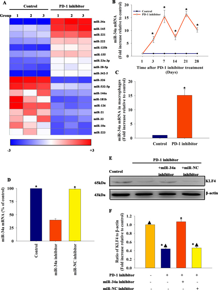
Fig. 2. MiR-34a was elicited when mice were treated with a PD-1 inhibitor.
a Heat map of miRs differentially expressed between mice treated with a PD-1 inhibitor and control mice, n = 3 per group. b MiR-34a expression was validated using qRT-PCR in mice treated with a PD-1 inhibitor and control mice. n = 6 per group *P < 0.05 versus the control group. c MiR-34a expression was validated using qRT-PCR in macrophages isolated from mice treated with a PD-1 inhibitor and control mice. n = 3 per group *P < 0.05 versus the control group. Mouse hearts were locally transfected with an inhibitor control (miR-NC inhibitor) or an miR-34a inhibitor. Untreated mice were used as a control. d Transfection efficiency was analyzed using qRT-PCR. n = 3 per group. *P < 0.05 versus miR-34a inhibitor. e, f Expression of KLF4 from hearts locally transfected with an inhibitor control (miR-NC inhibitor) or an miR-34a inhibitor in mice treated with a PD-1 inhibitor, only PD-1 inhibitor-treated mice, or untreated mouse heart tissue was examined by western blot analysis.compared with control mice n = 3 per group. *P < 0.05 versus the control group; ▲P < 0.05 versus the PD-1 inhibitor + miR-34a inhibitor group.

