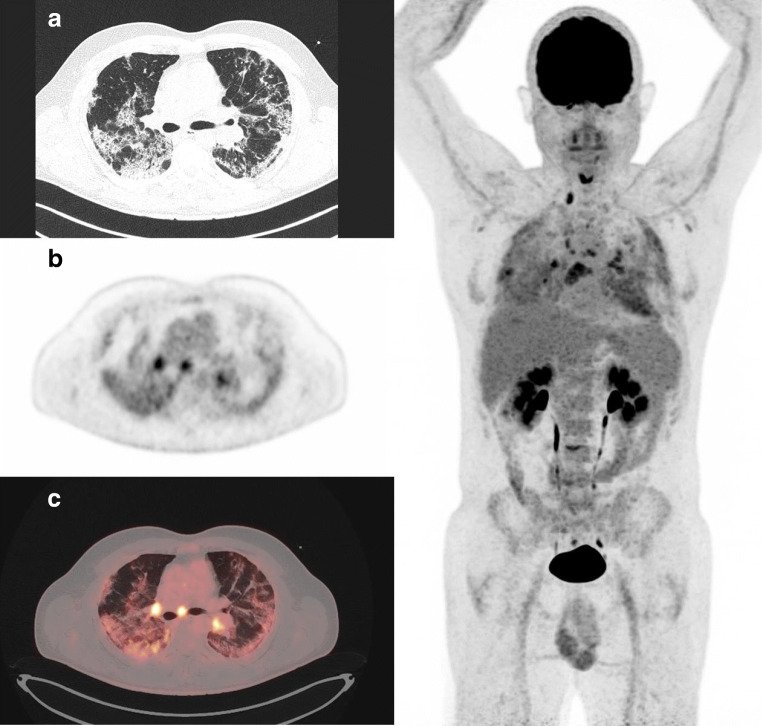
Fig. 3.
A 55-year-old man patient who presented severe respiratory symptoms in the days before FDG PET/CT. Images showed bilateral consolidation with mild increased FDG uptake (SUVmax 5). Despite this mild inflammation, the patient remained symptomatic for days requiring oxygen therapy through nasal canula at low flow rates and was discharged from hospital only 13 days later. Left: A CT transverse slice, B FDG PET slice, and C FDG PET and CT-fused images. Right: whole-body MIP image demonstrating that FDG uptake in mediastinal lymph nodes is above lung FDG uptake (respectively, SUVmax 6.9 versus 5)

