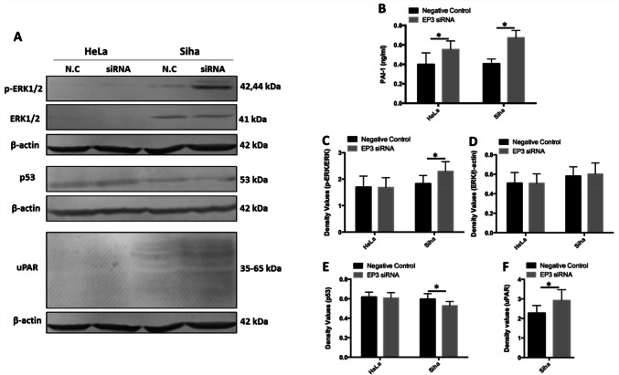
Fig. 4.
Expression of plasminogen activator inhibitor type 1 (PAI-1) and urokinase-type plasminogen activator receptor (uPAR) is influenced by silencing EP3 gene. a Western blotting analysis shows the expression of phosphorylated extracellular signal-regulated kinases (p-ERK1/2), extracellular signal-regulated kinases (ERK1/2), p53 and uPAR in HeLa and SiHa cells following treatment with EP3 siRNA and the negative control (N.C) for 48 h. β-actin was used as a loading control and all the data was normalized to the β-actin band signals. b PAI-1 levels in the supernatants of HeLa and SiHa cells are enhanced after silencing EP3 compared with the negative control for 48 h by ELISA (*P < 0.05, n = 6). c The histogram illustrates the expression of p-ERK1/2 is increased after silencing EP3 gene for 48 h in SiHa cells (*P < 0.05). d The histogram presents the expression of ERK1/2 is not altered by EP3 siRNA in HeLa and SiHa cells (P > 0.05). e The histogram illustrates the expression of p53 is inhibited after downregulation of EP3 compared with the negative control for 48 h in SiHa cells (*P < 0.05). f The histogram shows the expression of uPAR is stimulated after EP3 knockdown compared with the negative control for 48 h in SiHa cells (*P < 0.05). Statistically significant differences (P < 0.05) between EP3 siRNA group and the negative control group are marked with an *. All western blots data are shown as mean ± SD (n = 3). Full-length blots are shown in Supplementary Fig. 2

