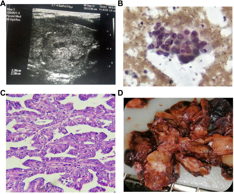
Figure 2.
A 32 years old female with papillary thyroid cancer (PTC), she was not diabetic or hypertensive, with negative family history for thyroid disease. She was euthyroid, Weight: 98 kg, Height: 164 cm, body mass index (BMI): 36.4, waist circumference (WC): 104 cm. (A) Ultrasonography showed the left thyroid nodule was 2.26 x 2.38 x 2.00 cm. The nodule was complex and isoechoic with eggshell calcifications, marked increase central/peripheral vascularity, incomplete halo, as well as irregular outline. (B) Cytopathology showed follicular cells with abundant cytoplasm, enlarged nuclei with vesicular chromatin, conspicuous eccentric nucleoli, focal nuclear overlapping and grooving (Pap stain, 400x original power). (C) Histopathology showed Papillae of malignant follicular cells with fibrovascular cores. The cells showed optically clear enlarged nuclei with overlapping and grooving (Hematoxylin and eosin stain, 100x original power). (D) Gross pathology showed a suspicious white firm thyroid nodule was detected grossly.

