
Figure 5.
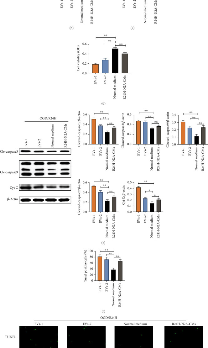
Effects of EVs derived from N2A cells on apoptosis of UC-MSCs upon OGD/R24H insult. UC-MSCs were precultured with two various concentration EVs, normal cultured medium, and R24H-N2A-CMs for 24 hours, respectively. Following treatment, UC-MSCs were subjected to OGD/R24H insult. (a–b) Apoptosis of UC-MSCs as evaluated by flow cytometry with Annexin-V/PI staining assay. (c) Apoptosis of UC-MSCs as evaluated by LDH leakage assay. (d) Cell viability of UC-MSCs as evaluated by MTT assay. (e) Expression and quantitative data of cleaved-caspase3, cleaved-caspase9, and cytochrome C as evaluated by western blotting. (f–g) Apoptosis of UC-MSCs as evaluated by TUNEL immunofluorescence. Scale bar = 50 μm. EVs-1: represented as higher concentration EVs recorded by NTA assay. EVs-2: represented as lower concentration EVs recorded by NTA assay. All data are presented as the mean value ± SD (n = 3). ∗p < 0.05; ∗∗p < 0.01, compared with normal culture medium group.