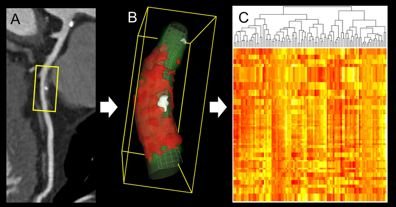
Figure 4. Radiomic analysis of plaque on coronary CTA.
(A) Curved multiplanar reconstruction of the left anterior descending artery showing mixed plaque in the midsegment. (B) For radiomic analysis, all voxels containing plaque are extracted from the region of interest. (C) Large numbers (typically >1,000) of intensity-, texture-, or shaped-based features are quantified to create big data, which can be analyzed using machine learning. CTA: computed tomography angiography. Reproduced with permission from Nicol et al. (120).

