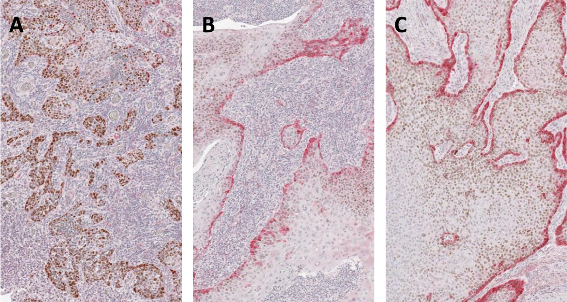
Fig. 1.

Quantification of p-EMT marker staining. Three cores from a single representative TMA with double IHC staining for p63 (brown) and a p-EMT marker (red). (A) Absent or minimal staining for red p-EMT marker (1+); (B) light or partial staining (2+); and (C) dark or complete staining (3+).
