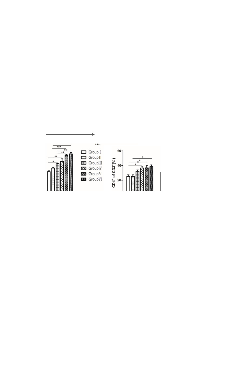
Fig. 5.
Lymphocyte proliferation and increased rates of CD3+CD4+ and CD3+CD8+ T cells. a Single cells were prepared from peripheral blood leukocytes (PBLs) as described, followed by stimulation with concanavalin A (10 μg/mL) for approximately 48 h. Evaluation of cell proliferation status by the MTT assay. b The percentage of CD4+ splenocytes was determined at 30 days and 38 days by FACS. c The percentage of CD8+ splenocytes was determined at 30 days and 38 days by FACS. (*P < 0.05, **P < 0.01 and ***P < 0.001)

