
Figure 3. Arginylation of BiP during treatment with ART, ERA, or BOR.
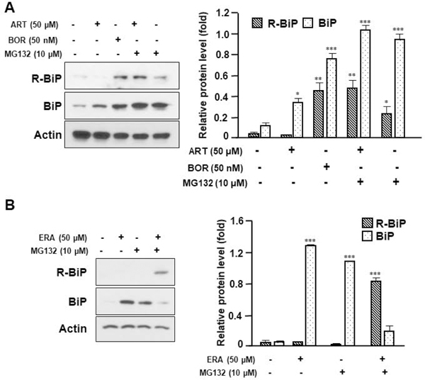
(A) HCT116 cells were treated with ART (50 μM) or BOR (50 nM) in the presence/absence of MG132 (10 μM) for 24 h. Western blotting analysis of BiP and arginylated BiP (R-BiP) was performed after treatment with indicated agents. Actin was used as a loading control. Densitometry analysis of the bands from the R-BiP or BiP was performed (right panel). The values are indicated as mean ± SD from three independent experiments. p-values: *, 0.05; **, 0.01; ***, 0.001. (B) HCT116 cells were treated with ERA (50 μM) in the presence/absence of MG132 (10 μM) for 24 h. Whole-cell extracts were analyzed with immunoblotting assay using indicated antibodies. Actin was used as a loading control. Densitometry analysis of the bands from the R-BiP or BiP was performed (right panel). The values are indicated as mean ± SD from three independent experiments. p-values: ***, 0.001.