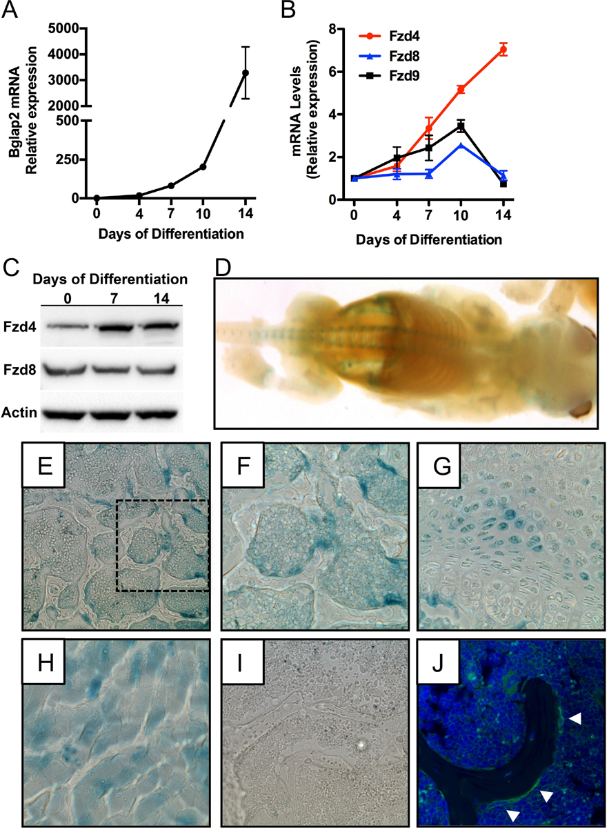
Figure 1. Fzd4 is highly expressed by osteoblasts.

(A and B) qPCR analysis of Bglap2 (A) and Fzd4, Fzd8, Fzd9 mRNA levels in primary osteoblasts cultured under osteogenic conditions for up to 14 days. (C) Western blot analysis of Fzd4 and Fzd8 protein levels in primary osteoblasts. (D) Whole mount X-gal staining of newborn (P0) Fzd4+/− mice that contain a lacZ reporter gene under the control of the endogenous promoter. (E-G) X-gal staining in the trabecular bone compartment (E and F) and growth plate (G) in the distal femur of 3 week old Fzd4+/− mice. 10X original magnification. Panel F shows a 20X magnification of the boxed section of panel E. (H and I) X-gal staining of skeletal muscle (H) from Fzd4+/− mice and distal femur (I) of Fzd4+/+ mice were used as a positive and negative control for staining, respectively). (J). Immunofluorescent staining for Fzd4 in the distal femur of a 3 week old mouse. White arrowheads indicated Fzd4 expressing osteoblasts.
