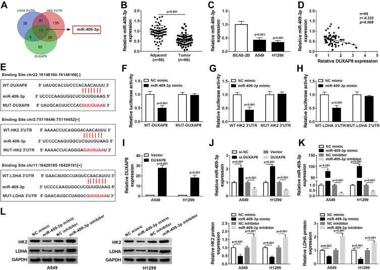
Figure 4.
DUXAP8 targeted miR-409-3p to regulate HK2 and LDHA expression. (A) The overlapped binding miRNAs were analyzed by Venn diagram. (B) The expression of miR-409-3p in NSCLC tissues was detected by RT-qPCR (n=3). (C) RT-qPCR was used to measure the expression of miR-409-3p in BEAS-2B, A549 and H1299 cells (n=3). (D) The relationship between DUXAP8 and miR-409-3p in NSCLC tissues was analyzed by Kaplan–Meier analysis (r = −0.322, P = 0.008) (n=3). (E) StarBase v2.0 predicted that miR-409-3p contained the binding sites of DUXAP8, HK2 and LDHA. (F and H) Dual-luciferase reporter assay was performed to determine the relative luciferase activity in 293T cells co-transfected with wild type (WT-) or mutant (MUT-) of DUXAP8, HK2 3ʹUTR or LDHA 3ʹUTR and miR-409-3p mimic or NC mimic (n=3). (I) The expression of DUXAP8 in NSCLC cells transfected with DUXAP3 or Vector was detected by RT-qPCR (n=3). (J) RT-qPCR was used to determine miR-409-3p expression in NSCLC cells transfected with si-NC, si-DUXAP8, Vector, or DUXAP8 (n=3). (K) RT-qPCR was used to detect the expression of miR-409-3p in NSCLC cells transfected with NC mimic, miR-409-3p mimic, NC inhibitor, or miR-409-3p inhibitor (n=3). (L) The protein expression of HK2 and LDHA in NSCLC cells transfected with NC mimic, miR-409-3p mimic, NC inhibitor, or miR-409-3p inhibitor was measured by Western blot (n=3).

