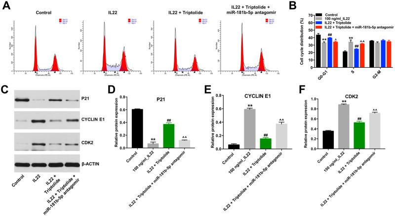
Figure 3.
Triptolide restrained the cell cycle in IL22-stimulated HaCaT via upregulating miR-181b-5p. HaCaT cells were transfected with miR-181b-5p antagomir and exposed to 10 μM triptolide for 24 h, and then treated with 100 ng/mL of IL22 for 24 h. (A and B) Cell cycle staging was measured by flow cytometry. (C) Expression levels of P21, CYCLIN E1 and CDK2 in cells were detected with Western blotting. (D–F) The relative expressions of P21, CYCLIN E1 and CDK2 in cells were quantified via normalization to BETAACTIN. **P < 0.01, compared with the control group. ##P < 0.01, compared with the 100 ng/mL IL22 group. ^P < 0.05, ^^P < 0.01 compared with IL22 + triptolide group.

