Fig. 1.
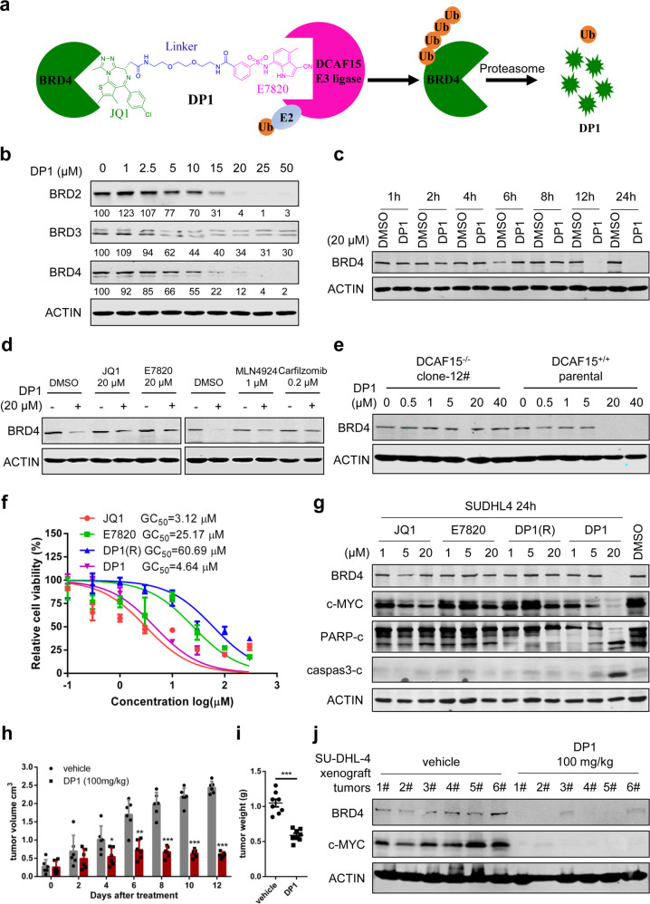
Design and characterization of DCAF15 E3 ligase derived PROTAC to degrade BRD4 in vitro and in vivo. a PROTACs designing schematic. b Immunoblot of BET protein and ACTIN after 16 h of treatment of SU-DHL-4 cells with the indicated concentrations of DP1. Degradation activity is calculated below each lane as % of protein level relative to DMSO control. c Immunoblot of BRD4 and ACTIN after treatment of SU-DHL-4 cells with 20 μM DP1 for the indicated incubation times. d Left panel: Immunoblot of BRD4 and ACTIN after a 4 h pretreatment with 20 μM of ligands JQ1 and E7820, followed with a 14 h 20 μM DP1 treatment in SU-DHL-4 cells. Right panel: Immunoblot of BRD4 and ACTIN after a 4 h pretreatment with carfilzomib (0.2 μM) or MLN4924 (1 μM), followed with a 14 h 20 μM DP1 treatment in SU-DHL-4 cells. e Immunoblot of BRD4 and ACTIN following treatment of clone 12 and parental cells for 24 h with the indicated concentrations of DP1. f Cell viability analysis of SU-DHL-4 cells treated with DP1 for 48 h compared with its component ligands JQ1, E7820, and DP1(R) (n = 3). g Immunoblot analysis of BRD4, c-MYC, cleaved PARP and Caspase 3, and ACTIN in SU-DHL-4 cells treated with the indicated concentrations of JQ1, E7820, DP1(R), and DP1 for 24 h. h Tumor volume of vehicle-treated mice or mice treated with DP1 (100 mg/kg) for 12 days, n = 8. i Tumor weight of mice treated with vehicle and DP1 after sacrificed. j Immunoblot of BRD4, c-MYC, and ACTIN using tumor lysates from mice treated with vehicle and DP1

