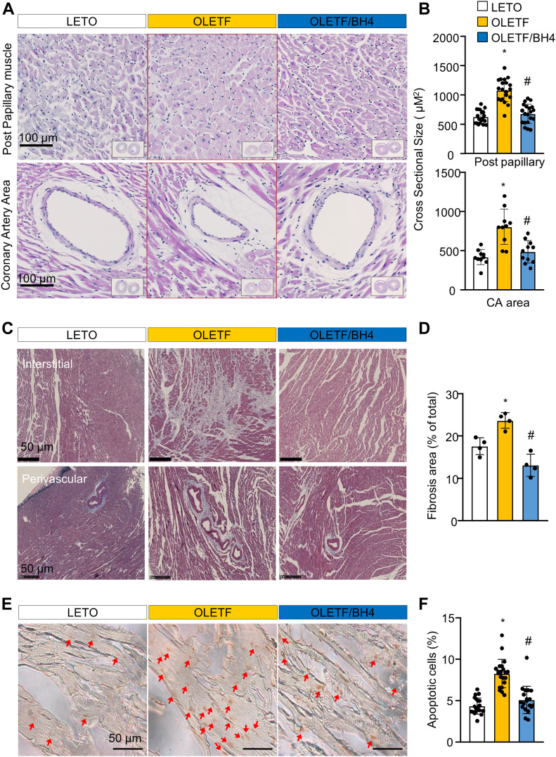
Figure 2. BH4 supplementation recovers pathological cardiac remodeling in a model of late-stage type 2 diabetes mellitus.
(A) Representative H&E staining of cross-sectioned hearts. (B) Cross-sectional size of cardiac muscle fibers. (C) Masson’s trichrome staining of cross-sectioned hearts. (D) Quantification of fibrotic area. (E) Images of TUNEL-stained heart tissues from experimental rats. Red arrow: TUNEL+ apoptotic cells. Scale bar: 50 μm. (F) Quantification of apoptotic cell death. Data in (A, B, C, D, E, F) represent the mean ± SEM (n = 4 animals/group). *P < 0.05 versus Long–Evans Tokushima Otsuka; #P < 0.05 versus Otsuka Long–Evans Tokushima Fatty. TUNEL, terminal deoxynucleotidyl transferase dUTP nick-end labeling.

