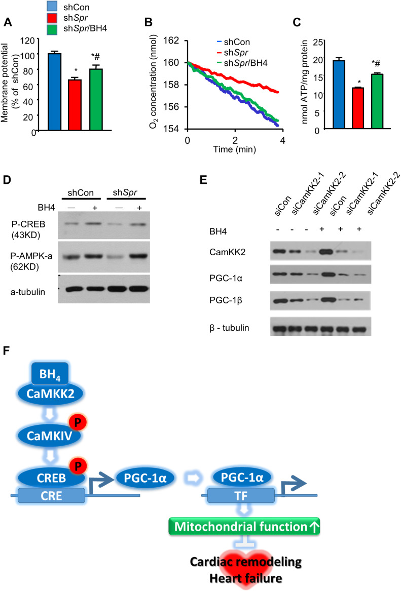
Figure 7. BH4 regulates PGC-1α levels by modulating CaMKK2 signaling.
(A) Relative mitochondrial membrane potentials. (B, C) Cellular oxygen consumption rates (B) and relative ATP levels (C) in indicated cell types. (D) Immunoblot analyses of p-CREB and p-AMPK-α in shCon, shSpr, and BH4 (20 μM)-treated shSpr HL-1 cells. (E) Representative Western blot of siCon and Camkk2-knockdown (siCaMKK2-1 or siCaMKK2-2) HL-1 cells in the presence or absence of BH4 (20 μM). (F) The proposed mechanism of BH4–CaMKK2-associated heart and mitochondrial regulation. *P < 0.05 versus shCon; #P < 0.05 versus shSpr (n = 3/group).
Source data are available for this figure.

