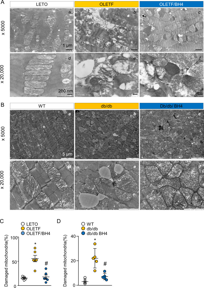
Figure S4. BH4 improves mitochondrial ultrastructure in diabetic rat and mouse hearts.
(A) Representative EM image of cardiac left ventricle tissue from Long–Evans Tokushima Otsuka (LETO), Otsuka Long–Evans Tokushima Fatty (OLETF), and OLETF/BH4 rats. (B) EM images of cardiac left ventricle tissue from WT, db/db, and db/db-BH4 mice. (C, D) Quantification of damaged mitochondrial proportion in hearts from LETO, OLETF, and OLETF/BH4 rats (C) and WT, db/db, and db/db-BH4 mice (n = 5/group). (C) *P < 0.05 versus LETO; #P < 0.05 versus OLETF. (D) *P < 0.05 versus WT; #P < 0.05 versus db/db.

