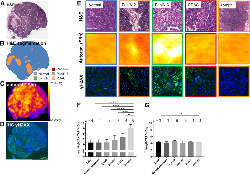
FIGURE 4.
(A) Hematoxylin and eosin staining of pancreas section from 113-d-old KPC mouse harvested 24 h after administration of 111In-anti-γH2AX-TAT. (B) Identification of various morphopathologic features. (C) Autoradiography image showing distribution of radioactivity. (D) Immunofluorescence image showing γH2AX (green) and nuclei (blue). Additional sections are shown in Supplemental Figure 6. (E) Magnification of histologic areas in A–D. (F) Uptake of 111In-anti-γH2AX-TAT in various morphopathologic features in KPC pancreata, measured by ex vivo autoradiography of pancreas sections. (G) Uptake of 111In-IgG-TAT in various morphopathologic features in KPC pancreata, measured by ex vivo autoradiography of pancreas sections. Autorad = autoradiography; H&E = hematoxylin and eosin; IHC = immunohistochemistry; lymph = lymphocytes.

