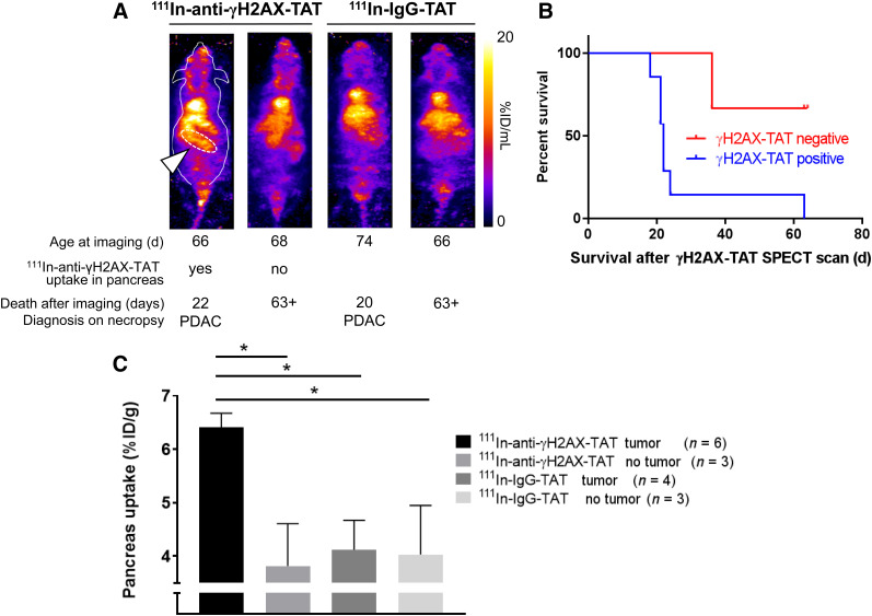
FIGURE 5.
(A) Representative images of KPC mice aged 66–77 d imaged by SPECT, 24 h after intravenous administration of 111In-anti-γH2AX-TAT. Age at time of imaging, length of survival before clinical symptom endpoints were reached, and diagnosis at necropsy are indicated for each mouse. Pancreatic region is indicated by white arrowhead and white dashed line in first animal only. Coronal maximum-intensity projections are shown; outline of mouse is indicated for first animal only. (B) Mice showing uptake of 111In-anti-γH2AX-TAT in pancreas had significantly shorter survival than those not showing pancreatic uptake (P = 0.0273). (C) 111In-anti-γH2AX-TAT, but not 111In-IgG-TAT, was taken up more in pancreata of tumor-bearing KPC mice.

