Fig. 3.

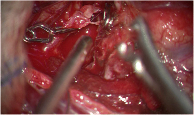
Intraoperative microscopic image is showing the pseudoaneurysm of the M2 segment of the left middle cerebral artery. Besides, the dissection is shown along the length of the affected artery

Intraoperative microscopic image is showing the pseudoaneurysm of the M2 segment of the left middle cerebral artery. Besides, the dissection is shown along the length of the affected artery