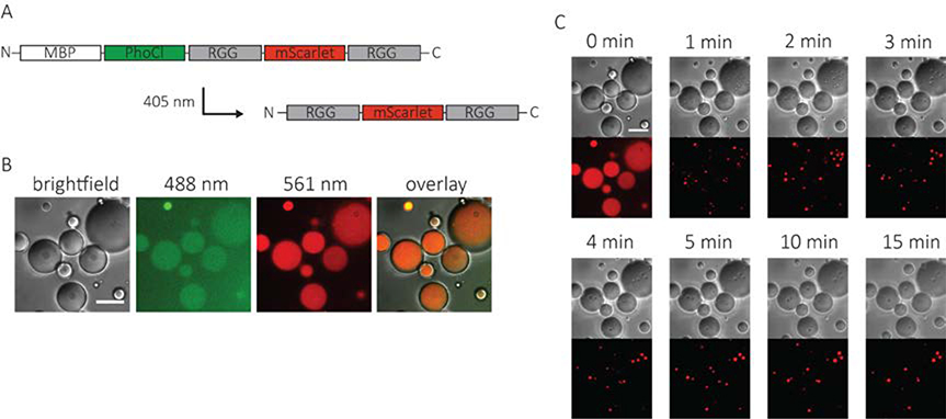
Figure 3. Incorporation of a folded protein into optically-regulated protein coacervates.
A. Schematic of an optically active RGG construct containing mScarlet. B. Images of water-in-oil emulsions containing 2 mg/mL MBP-PhoCl-RGG-mScarlet-RGG prior to exposure to 405 nm light. The aqueous phase contained 150 mM NaCl and 20 mM tris, pH 7.4. Image was taken in DIC, 488 nm, and 561 nm channels. C. Time-lapse DIC and 561 nm channel images of protein-containing emulsions after exposure to 405 nm light (5 second light pulse, 7.9 mW/cm2). Scale bars: 20 μm. Image intensities were background adjusted for clarity.

