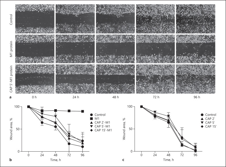
Fig. 1.
M1 protein-triggered inhibition of cell recovery processes is abolished upon CAP treatment. a Representative cell images of the ability of keratinocytes to heal a scratch after treatment with M1 protein (5 μg/mL), CAP-treated M1 protein (5 μg/mL), or left untreated, followed over 96 h. After 48 h, cell culture media were exchanged without addition/supplementation of bacterial protein. Cell images were monitored every 24 h and wound areas were quantified by the ImageJ software utilising the macro MRI Wound Healing Tool. b Summary of scratch assays supplemented with M1 protein and CAP-treated M1 protein at indicated time points and HaCaT cells added (c) with CAP-treated cell culture media without bacterial protein at indicated time points. Untreated cells served as control. All results were analysed by two-way ANOVA and Tukey's multiple comparisons test, resulting in a significant difference (p < 0.01) of M1 protein-treated cells compared to control after 48 h as well as compared to all groups (p < 0.0001) after 72 and 96 h. The remaining comparisons were statistically insignificant. The data represent the average percentage of initial wound area ± SD from three independent experiments for every group. CAP, cold atmospheric plasma.

