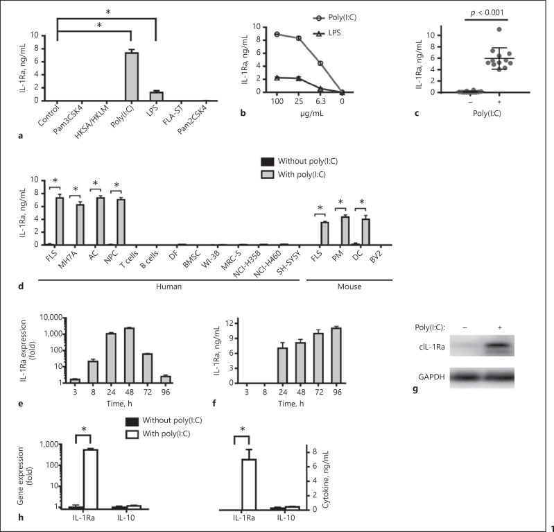
Fig. 1.
Poly(I:C) induces IL-1Ra expression in several types of cells. Human FLS (105 cells/mL) were stimulated with the indicated TLR agonists, including Pam3CSK4, HKSA/HKLM, poly(I:C), LPS, FLA-ST, or Pam2CSK4 for 24 h (a). Human FLS were stimulated by poly(I:C; 100, 25, 6.3 μg/mL) or LPS (100, 25, 6.3 μg/mL) for 24 h (b). Human FLS from 12 individuals with RA, femoral head necrosis or fracture were stimulated by poly(I:C) for 24 h (c). Human FLS (105 cells/mL), MH7A (105 cells/mL), AC (5 × 10⁵ cells/mL), NPC (2 × 10⁵ cells/mL), T cells (106 cells/mL), B cells (106 cells/mL), DF (5 × 10⁴ cells/mL), BMSC (5 × 10⁴ cells/mL), WI-38 cells (105 cells/mL), MRC-5 cells (2.5 × 10⁵ cells/mL), NCI-H358 cells (4 × 10⁵ cells/mL), NCI-H460 cells (4 × 10⁵ cells/mL) and SH-SY5Y cells (5 × 10⁵ cells/mL), and mouse FLS (105 cells/mL), PM (5 × 10⁵ cells/mL), DC (5 × 10⁵ cells/mL) and BV2 (5 × 10⁵ cells/mL) were stimulated by poly(I:C) for 24 h (d). Human FLS were stimulated by poly(I:C) for 3, 8, 24, 48, 72 or 96 h. The cells were harvested and the levels of IL-1Ra mRNA expression were measured by quantitative real-time PCR (e). Upregulation of IL-1Ra expression is compared with unstimulated cultures. The concentrations of IL-1Ra in the culture supernatants were measured by ELISA (f). The cells were harvested and the relative levels of cytoplasmic IL-1Ra expression were assessed by Western blot (g). Human FLS were stimulated by poly(I:C) for 24 h (h). The cells were harvested and the levels of IL-1Ra and IL-10 mRNA expression were measured by quantitative real-time PCR. Upregulation of IL-1Ra and IL-10 expression is compared with unstimulated cultures. The concentrations of IL-1Ra and IL-10 in the culture supernatants were measured by ELISA. Results are presented as mean ± SEM (n = 5 per group in a, b, d–f, n = 12 per group in c) from 3 separate experiments. * p < 0.05 versus the group without poly(I:C). IL, interleukin; IL-1Ra, IL-1 receptor antagonist; LPS, lipopolysaccharides; AC, articular chondrocytes; NPC, nucleus pulposus cells; PM, peritoneal macrophages; DC, dendritic cells; FLS, fibroblast-like synoviocytes; DF, dermal fibroblasts; BMSC, bone marrow mesenchymal stem cells.

