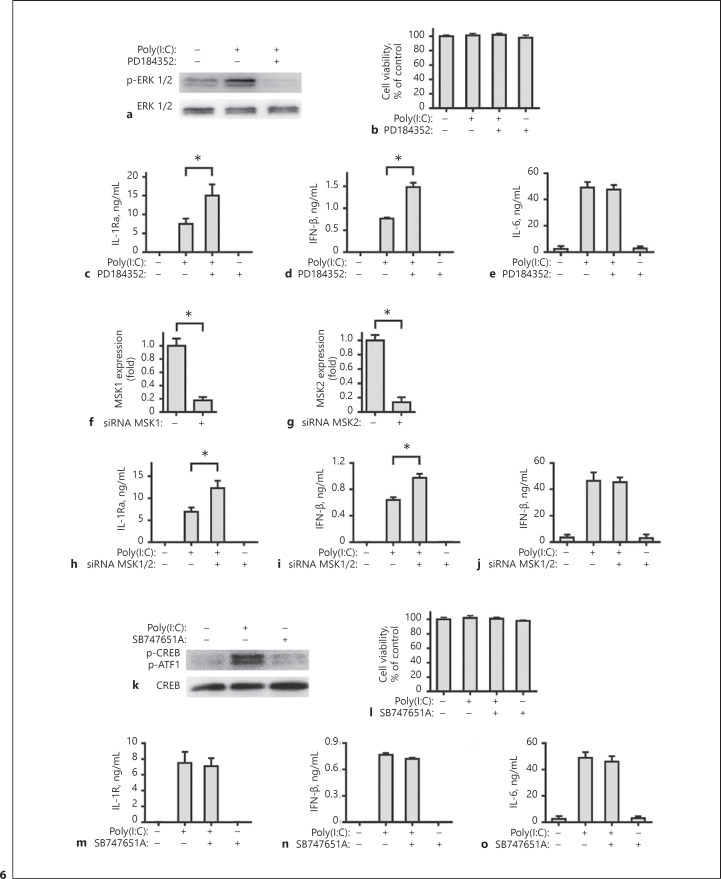
Fig. 6.
Induction of IL-1Ra expression by TLR3 activation is negatively regulated by the ERK-MSKs signaling. Human FLS were pre-treated with PD184352 (2 μM) or SB747651A (10 μM) for 1 h. Subsequently, the cells were stimulated with poly(I:C) for 30 min and the relative levels of ERK 1/2 (a) and CREB (k) expression and phosphorylation in the different groups of FLSs were measured by Western blot assays. Human FLS were pre-treated with PD184352 (2 μM, b–e) or SB747651A (10 μM, l–o) for 1 h and stimulated by poly(I:C) for 8 h or 24 h. The cell viability was measured by CCK-8 assay. The concentrations of IL-1Ra, IFN-β and IL-6 in the culture supernatants were determined with ELISA. Human FLS were treated with siRNA MSK1 (f), siRNA MSK2 (g), or siRNA NC for 48 h. The cells were harvested and the levels of human MSK1 and MSK2 mRNA expression were measured by real-time PCR. Downregulation of human MSK1 and MSK2 expression are compared with the siRNA NC group. Human FLS were pre-treated with siRNA MSK1 and MSK2, or siRNA NC for 48 h and stimulated by poly(I:C) for 8 h or 24 h. The concentrations of IL-1Ra (h), IFN-β (i), and IL-6 (j) in the culture supernatants were determined with ELISA. Results are presented as mean ± SEM (n = 5 per group) from 3 separate experiments. * p < 0.05 vs. the group with poly(I:C) and vehicle. IL, interleukin; IL-1Ra, IL-1 receptor antagonist.

