Fig. 1.
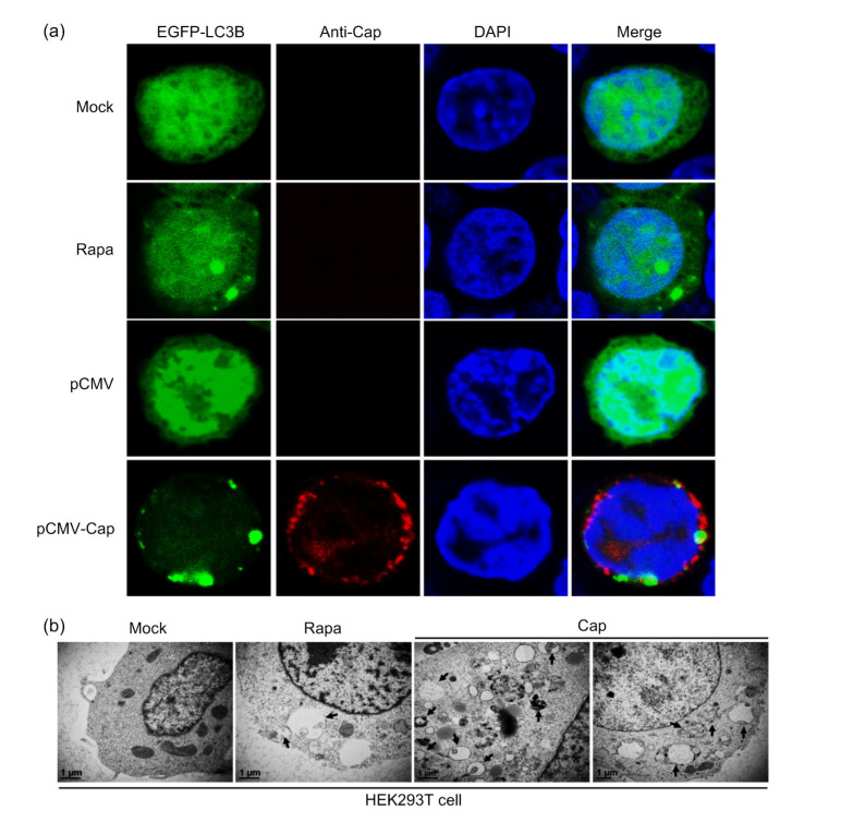
Formation of autophagosomes induced by PCV3 capsid protein shown as EGFP-LC3B punctae and autophagosome-like vesicles
(a) Cells were co-transfected with pEGFP-LC3B and pCMV-capsid (Cap) or the control plasmid pCMV, or transfected only with pEGFP-LC3B (mock), or treated with 2 μmol/L rapamycin (Rapa) after 5 h of transfection with pEGFP-LC3B (positive control). After 36 h of transfection, cells were immuno-stained with the rabbit anti-PCV3 Cap polyclonal antibody and counterstained with 2-(4-amidinophenyl)-6-indolecarbamidine dihydrochloride (DAPI) for nuclei, and then examined for formation of EGFP-LC3B punctae under the confocal microscope. (b) Cells were transfected with pCMV-Cap for 36 h or mock-treated (negative control) or treated with 2 μmol/L Rapa (positive control). Autophagosome-like vesicles were observed by transmission electron microscope. Black arrows indicate the characteristic structures of autophagosomes. Scale bar=1 μm

