Figure 2.

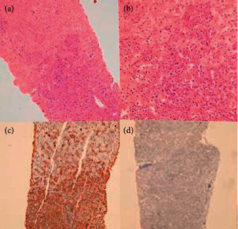
(a, b) Liver paranchyme is infiltrated by plasma cells and full of atypical cells. (c) Kappa (+) neoplastic plasma cells infiltrations are seen. (d) Lambda (−) neoplastic plasma cells infiltrations.

(a, b) Liver paranchyme is infiltrated by plasma cells and full of atypical cells. (c) Kappa (+) neoplastic plasma cells infiltrations are seen. (d) Lambda (−) neoplastic plasma cells infiltrations.