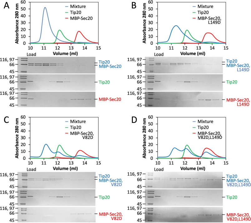
Figure 3.
Structure-guided disruption of S. cerevisiae Tip20•Sec20 complex formation. A, size-exclusion chromatography demonstrates robust binding (blue) between WT S. cerevisiae Tip20 (green) and MBP-Sec20NTD (red). B, substitution of Sec20 Leu-149 with aspartate has little effect on complex formation. C, substitution of Sec20 Val-82 with aspartate partially compromises binding to Tip20. D, the two substitutions in combination have a stronger effect on binding. The data presented for Tip20 alone in B–D are identical to those in A.

