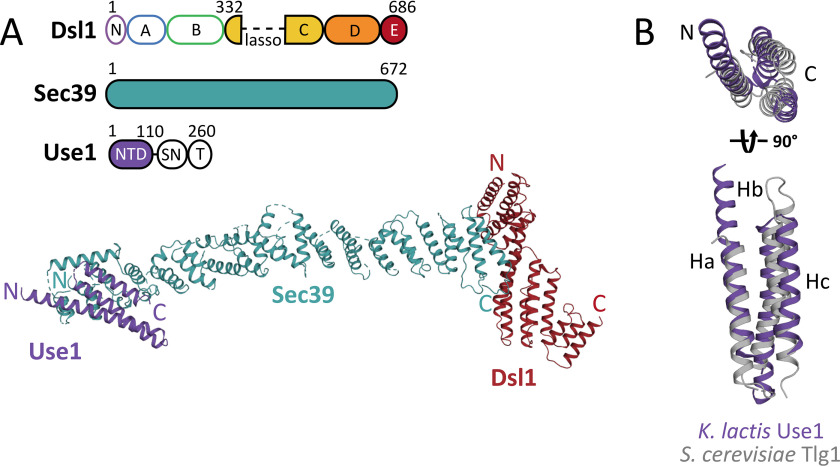
Figure 5.
6.5 Å-resolution X-ray structure of K. lactis Sec39•Use1NTD•Dsl1C–E. A, the upper panel depicts the domain architecture of the three proteins crystallized. Dsl1 consists of an N-terminal Tip20-interacting domain (N) followed by five CATCHR domains (A–E). Sec39 consists of a long α-solenoid. For further details, see Fig. S6. The SNARE protein Use1 consists of an N-terminal domain (NTD), a SNARE motif (SN), and a transmembrane helix (T). In the lower panel, Use1 (purple) binds to the extreme N terminus of Sec39 (blue). B, K. lactis Use1 (purple) superimposes on its closest homologue, S. cerevisiae Tlg1 (gray, PDB accession code 2C5K, chain T) with an root-mean-square deviation of 4.3 Å over 75 residues of the structure.

