Figure 4.
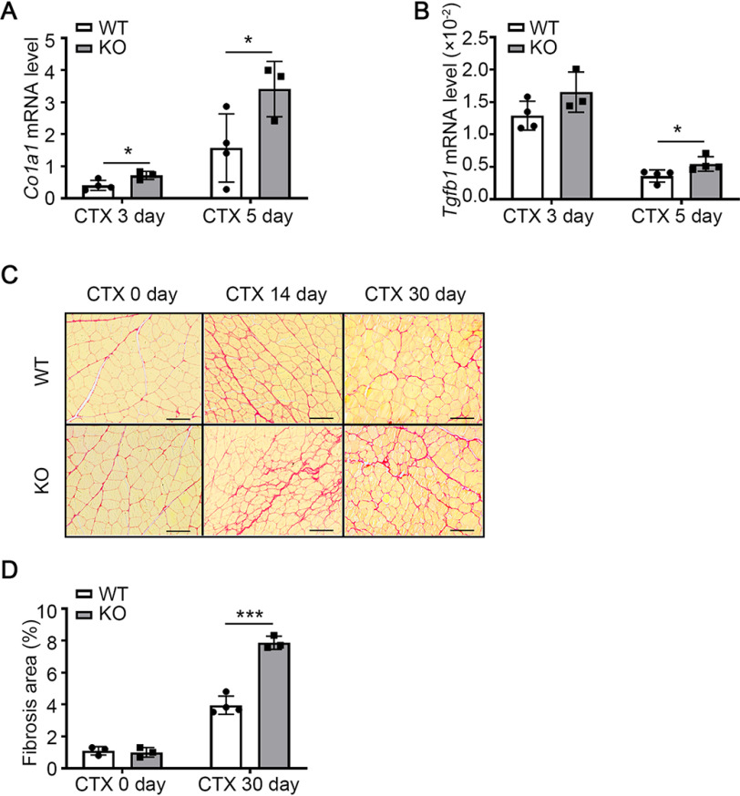
miR-223-3p KO promotes interstitial fibrosis in injured skeletal muscle. A–B, RT-PCR assessment of Col1a1 and Tgfb1 expression in the muscles of WT and miR-223-3p KO mice 3 days and 5 days after CTX injury (n = 3–4 per group). C, representative Picrosirius red staining (red) of muscles of WT and miR-223-3p KO mice on 0 day and 30 days after CTX injury. Scale bar, 100 μm. D, percentages of Picrosirius red-positive areas per field in the muscles of WT and miR-223-3p KO mice on 0 day and 14 days after CTX injury (n = 3–4 per group). Data are expressed as the mean ± S.D. *, p < 0.05; ***, p < 0.001 by unpaired two-tailed Student's t test.

