
Figure 1.
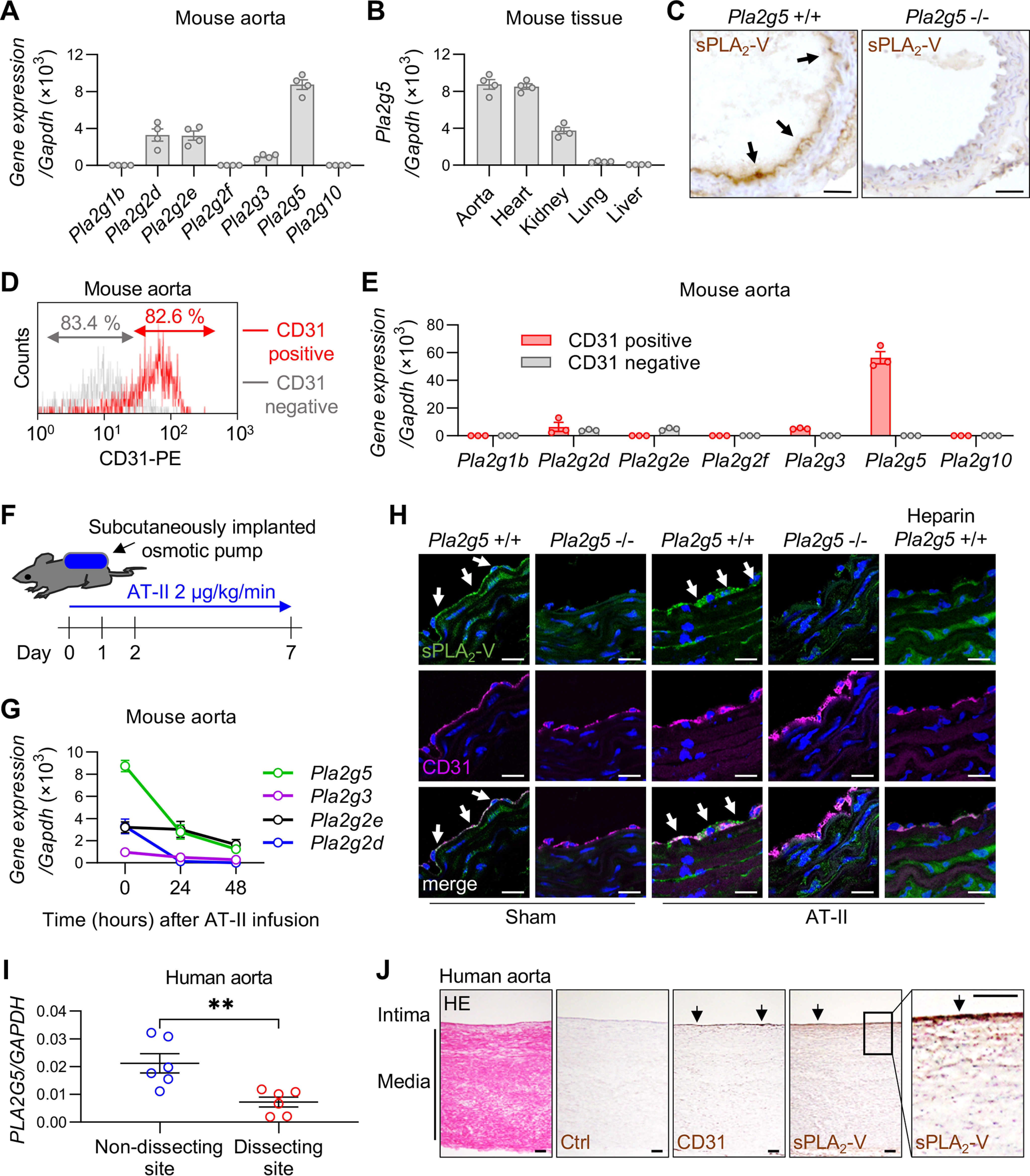
Expression of sPLA2s in the aortas of mice and humans. A, expression of sPLA2 mRNAs relative to Gapdh in the aorta of WT C57BL/6 mice (n = 4). B, expression of Pla2g5 in various tissues of WT mice (n = 4). C, immunohistochemistry of sPLA2-V in the aorta of Pla2g5+/+ and Pla2g5−/− mice. Arrows, positive staining of sPLA2-V. Scale bars, 100 μm. D, flow cytometry of CD31-positive/negative cells from WT mouse aorta. E, expression of sPLA2 mRNAs of CD31 positive/negative cells from WT mouse aorta (n = 3). F, schematic procedure of AT-II infusion into mice using subcutaneously implanted osmotic pumps. G, time course of the expression of sPLA2 mRNAs in WT mouse aorta after AT-II infusion (n = 4). H, immunofluorescence of sPLA2-V (green) and CD31 (magenta) with DAPI (blue) in the aorta of Pla2g5+/+ and Pla2g5−/− mice with or without AT-II infusion for 48 h. The rightmost panels represent the staining of Pla2g5+/+ aorta after AT-II infusion, following perfusion with heparinized saline through the left ventricle before extraction. Arrows indicate positive staining of sPLA2-V. Scale bars, 20 μm. I, expression of PLA2G5 in nondissecting or dissecting site of human aorta (n = 6). **, p < 0.01 by unpaired t test. J, a representative section of human aorta stained with HE and immunohistochemistry of the serial sections with control IgG (Ctrl), anti-CD31 antibody, or anti-sPLA2-V antibody. A boxed area is magnified on the right. Arrows, positive staining. Scale bars, 100 μm. Data are presented as mean ± S.E. (error bars) of the indicated number (n) of biological replicates.