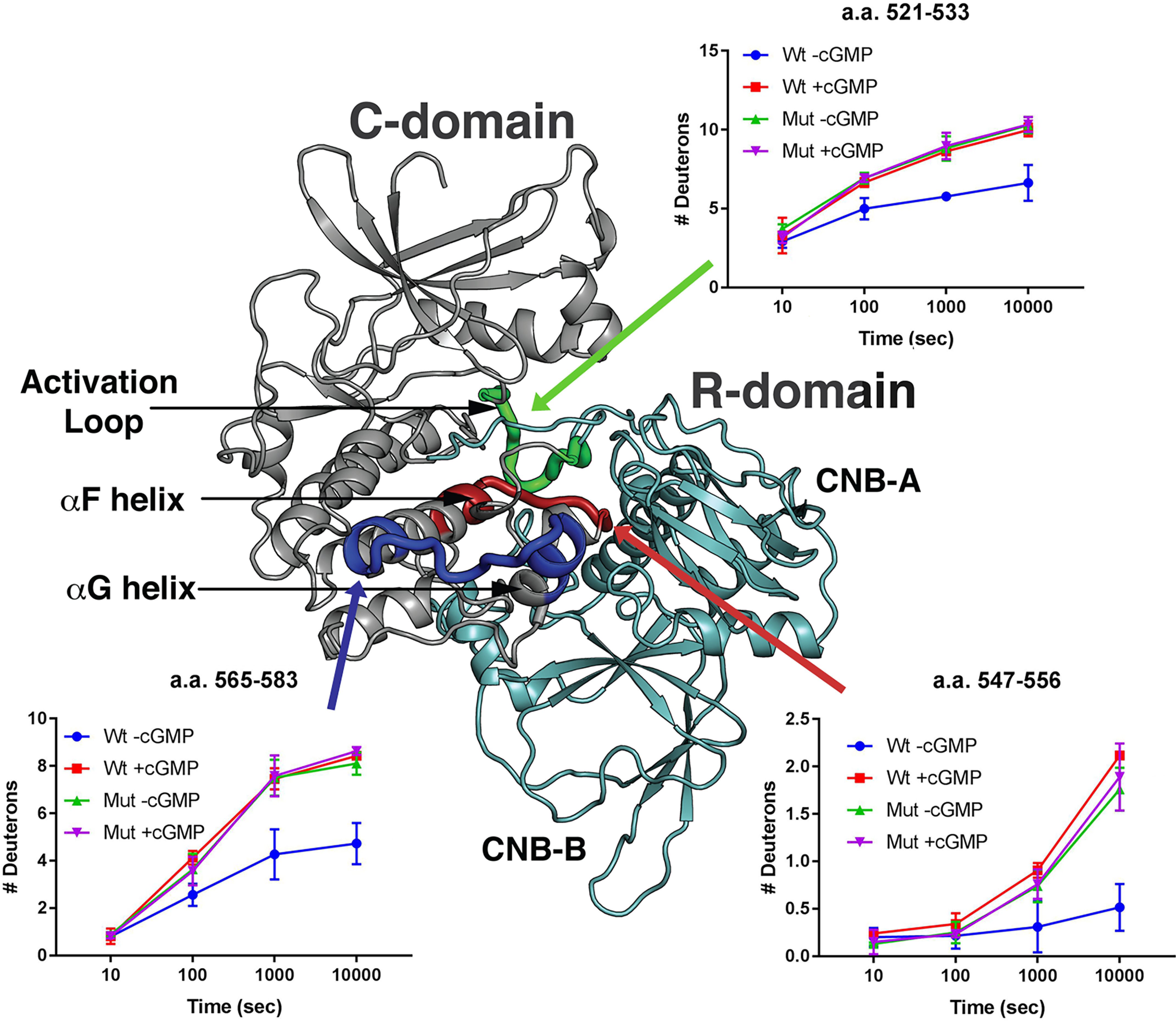
Figure 8.

H/D exchange in the catalytic cleft of WT and R192Q PKG1β. H/D exchange profiles of peptides from WT (Wt) and R192Q (Mut) PKG1β in the presence or absence of cGMP are shown mapped to a molecular model of inactive PKG1β. The regulatory domain is colored teal and the catalytic domain is mainly colored gray, with the following exceptions: the region containing amino acids 521–533 is colored green; 547–556 is colored red; and 565–583 is colored dark blue. Graphs show the number of deuterons incorporated into the peptides as a function of time. H/D exchange data are the averages from two independent H/D exchange reactions performed with separate protein preparations.
