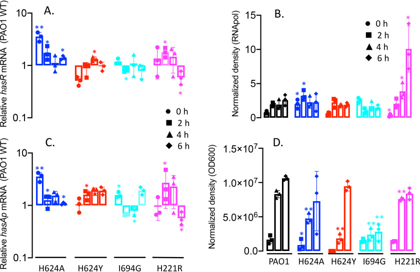
Figure 3.
Relative HasR and HasAp mRNA and protein levels for the hasR allelic strains under iron-depleted conditions. A, hasR mRNA isolated at 0, 2, 4, and 6 h following growth in M9 minimal media. mRNA values represent the mean from three biological experiments, each performed in triplicate and normalized to PAO1 WT at the same time point. Error bars represent the standard deviations from three independent experiments performed in triplicate. The indicated p values were normalized to mRNA levels of PAO1 WT at the same time point: *, p < 0.05. B, Western blot analysis of PAO1 WT and the hasR allelic strains. For HasR, total protein (5 μg) was loaded in each well. RNApolα was used as a loading control. Normalized density (n = 3) was performed for three separate biological replicates. The indicated p values were normalized to PAO1 at the same time point: *, p < 0.05; **, p < 0.005. C, hasAp mRNA analyzed as in panel A. D, Western blot analysis of HasAp as in panel B. Extracellular supernatant (4 μl) was loaded and run on the automated Wes capillary Western system as described in Experimental procedures. Representative Western blot images are shown in the supporting information (Fig. S1).

