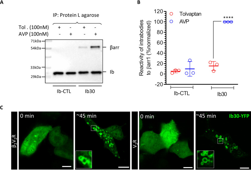
Figure 6.
Intrabody30 recognizes receptor-bound endogenous βarr1 and reports the trafficking of native βarr1. A, the ability of intrabody Ib30 to recognize V2R-bound endogenous βarr1 upon agonist stimulation. HEK-293 cells expressing V2R and HA-tagged Ib30/Ib–CTL were stimulated with either inverse agonist (tolvaptan; 100 nm) or agonist (AVP; 100 nm) followed by co-IP using anti-HA antibody agarose. Subsequently, the proteins were visualized by Western blotting using anti-βarr and anti-HA antibodies. B, densitometry-based quantification of the data in A presented as means ± S.E. from three independent experiments normalized with maximal response (treated as 100%) and analyzed using one-way ANOVA. ****, p < 0.0001. C, HEK-293 cells expressing β2V2R/V2R and Ib30–YFP were stimulated with isoproterenol (10 μm) and AVP (100 nm), respectively, and the localization of Ib30–YFP was visualized using confocal microscopy. Representative images from three independent experiments are shown here. Scale bar, 10 μm.

