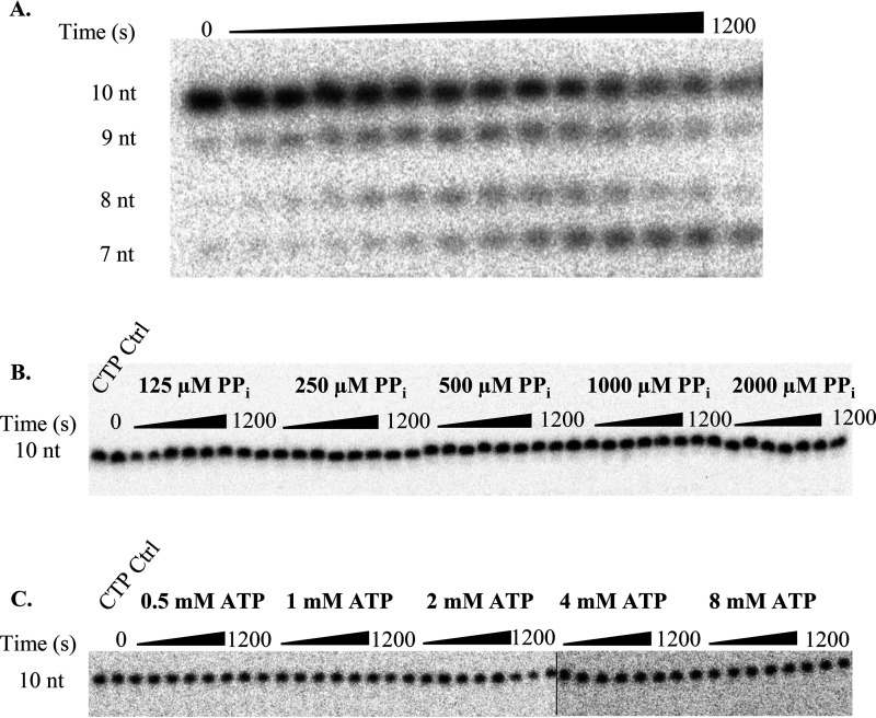
Figure 7.
Excision of 2′C-Me-2′F-UTP. A, representative 16% denaturing PAGE separation of UMP pyrophosphorolysis. Bands below the 9 nt band appear due to pyrophosphorolysis occurring at that position. All bands below 10 nucleotides were summed during analysis to obtain the correct concentration of the loss of the 10-nt substrate due to pyrophosphorolysis. B and C, results for pyrophosphorolysis of 2′C-Me-2′F-UMP (B) and ATP-mediated 2′C-Me-2′F-UMP excision (C). Reactions were separated on a 16% polyacrylamide gel containing 8 m urea. The line in B indicates where gels are spliced together. No excision products were detectable during the observed time course. The addition of the next correct nucleotide did not result in extension of the primer strand to an 11-nt product (CTP Ctrl, first lane in B and C), indicating effective chain termination.

