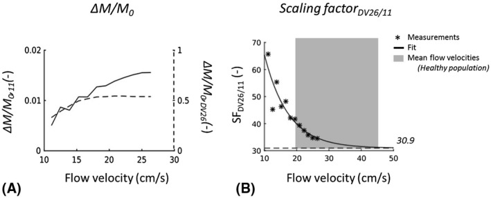
FIGURE 1.

A, ΔM/M 0 plotted against the flow velocity set for the flow phantom for the pCASL implementation at software version 11 (solid line, left y‐axis) and for the implementation at DV26 (dashed line, right y‐axis). B, Scaling factor (SF) between ΔM/M 0 within perfusion ROI of the phantom collected with pCASL sequences at DV26 and at 11. SF is dependent on the flow velocity of the perfusate at the location of labeling, with a fitted asymptote of 30.9 (close to GE’s scaling factor of 32, used in image recon at DV26). The gray box indicates the range of mean flow velocities in the internal carotid and vertebral arteries in a healthy population (N = 180, 20‐79 years), measured with Doppler sonography 17
