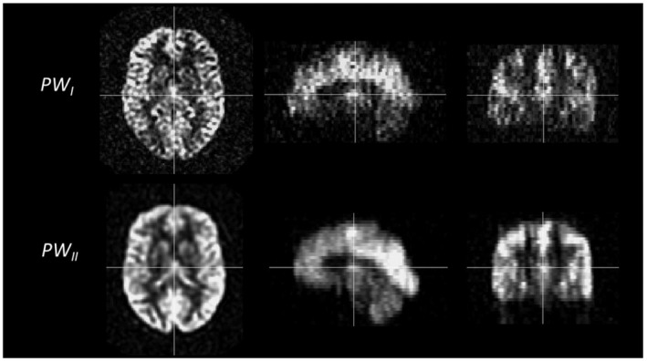
FIGURE 2.

Example of perfusion weighted (ΔM) images of one young volunteer. Note the difference in smoothing occurring during the reconstruction of the images. On scanner I with software version 11 (top row), no smoothing is implemented during reconstruction. On scanner II with software version DV26 (bottom row), smoothing occurs during reconstruction of the images
