Figure 3.

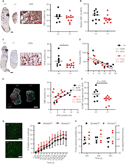
von Willebrand factor (VWF) and platelet characteristics of SLC44A2 deficient mice following 48 hours stenosis and ex vivo under flow conditions. A, Immunohistochemical (IHC) staining of VWF in thrombus of a representative wild type (Slc44a2+ / +, +/+) and SLC44A2 deficient mice (Slc44a2− / −, −/−), with high magnification from Slc44a2+ / + (location is indicated by the black box; left) and quantification (right) of positive area as a percentage of total thrombus area in Slc44a2+ / + (n = 8) or Slc44a2− / − mice (n = 11). B, Plasma VWF levels after 48 hours stenosis expressed as a percentage of MMP (mouse pool plasma; +/+ n = 9, −/− n = 12). C, IHC staining of glycoprotein Ib (GPIb) in thrombus of a representative Slc44a2+ / + and Slc44a2− / −, with high magnification from Slc44a2+ / + (location is indicated by the black box; left) and quantification (right) of positive area as a percentage of total thrombus area (+/+ n = 8, −/− n = 11). D, Correlation plot between thrombus weight and circulating blood platelets (+/+ n = 9, −/− n = 12). E, Immunofluorescent co‐stain of GPIb (red) and VWF (cyan) on thrombus sections (left) with correlation plot (right; n = 8 +/+; 11 −/−). F, Percentage of VWF positive area colocalized with GPIb. G, Representative images of DiOC6‐labeled platelets of a wild type (Slc44a2+/+, +/+) control (up) and a SLC44a2 deficient mouse (Slc44a2− / −, −/−; down) on a surface coated with VWF‐binding peptide. Scale bar is 20 µm. H, Percentage of stable platelet area coverage over a 30 second time period in field view within heparinized and D‐phenylalanyl‐prolyl‐arginyl chloromethyl ketone (PPACK) treated whole blood flowing over slides coated with a murine VWF binding peptide at venous shear rate (150 s−1) over time (n = 5 per group). (Note on numbers: blood samples that became coagulated or thrombi that were damaged during sectioning were not included for subsequent analysis.) I, Percentage platelets displaying a firm or transient interaction with VWF‐binding peptide determined by a method described by Meyer dos Santos et al. 17 Black and white bars equal 500 µm. Red bar equals 50 µm. Statistical differences between for platelet perfusion were evaluated by two‐way analysis of variance and for remaining biological readouts, the Mann‐Whitney rank‐sum test. Coefficient r calculated using Spearman's correlation (*signifies P < .05)
