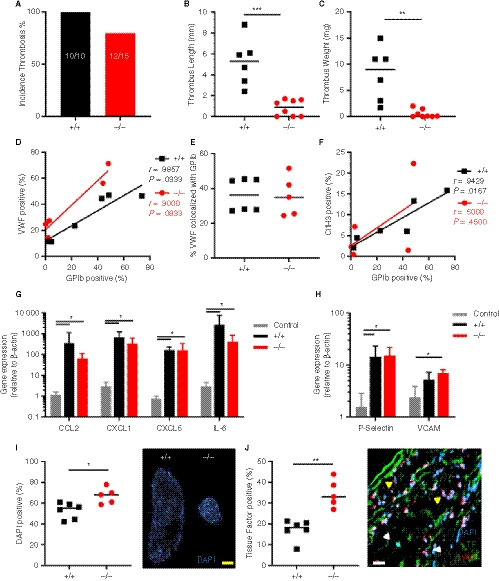
Figure 4.

Thrombosis in SLC44A2 deficient mice following 6 hours stenosis of the inferior vena cava (IVC). A, Incidence of thrombosis after 6 hours in mice wild type (+/+, n = 6) or SLC44A2 deficient mice (−/−, n = 8), shown as percentage. B, Length and (C) weight of thrombi formed at 6 hours. D, Correlation plot of GPIb and VWF immunostaining on thrombus sections (+/+, n = 6; −/−, n = 5). E, Percentage of VWF positive area colocalized with GPIb. F, Correlation plot of GPIb and citrullinated histone H3 immunostaining on thrombus sections. G, Transcript levels of inflammatory molecules produced by the local IVC in Slc44a2+ / + (+/+, n = 6) or Slc44a2− / − (−/−, n = 5) for SLC44A2 collected at 6 hours post‐stenosis as compared to non‐ligated control C57BL/6J mice (n = 3); chemokine (C‐C motif) ligand 2 (Ccl2), chemokine (C‐X‐C motif) ligand 1 (Cxcl1), Cxcl5, interleukin‐6 (Il6). H, IVC expression of mRNA transcripts encoding adhesion molecules; P‐Selectin and vascular cell adhesion molecule 1 (VCAM1) following 6 hours stenosis. Bars represent mean values with standard deviation (error bars). I, Nuclear stain (DAPI) of leukocytes in thrombi from Slc44a2+ / + and Slc44a2− / − mice (top left) with quantification as a percentage of total thrombus area (n = 6 +/+; 5 −/−). J, Quantification of immunofluorescent (IF) staining of tissue factor (TF; left) as a percentage of total thrombus area. Representative image of IF co‐stain by confocal microscopy of neutrophil marker Ly6G, TF, and nuclear stain (DAPI; right panel). Yellow arrows indicate Ly6G positive cells and white arrows indicate TF positive cells. Yellow bar equals 500 µm and white bar equals 20 µm. Statistical differences were evaluated using Mann‐Whitney rank‐sum test (*signifies P < .05; **signifies P < .01; ***signifies P < .001)
