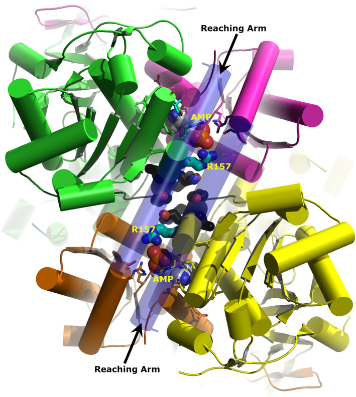
Figure 5.

Model of LiPFK based on the X‐ray structure of Trypanosoma brucei PFK (TbPFK). The view is along the twofold axis relating Dimer 1 to Dimer 2. Dimer 1 is coloured green and magenta and Dimer 2 orange and yellow. AMP is shown as spheres within each monomer–monomer interface. Arg157 is shown as thick sticks with cyan carbons. Residues adjacent to Arg157 and found within the dimer–dimer interface are shown as thick sticks with black carbons. The long helical ‘reaching’ arms are shown as slightly transparent and form a lid over the AMP sites and stretch across the dimer–dimer interface. Graphic created with The PyMOL Molecular Graphics System (version 1.8.6.2, Schrödinger, LLC).
