Figure 7.
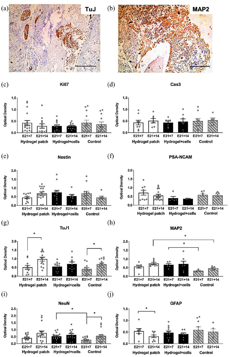
Quantitative histopathologic analysis of myelomeningocele organotypic slice cultures after hydrogel patch treatment (with or without neural progenitors) for 7 days (E21 + 7) or 14 days (E21 + 14). (a) and (b) Representative appearance of slice culture sections after staining using antibodies against TuJ1 and MAP2, respectively. Scale bar represents 500 mm. (c) and (d) Ki67 and Cas3 as markers of cell proliferation and apoptosis, respectively. Control slice cultures in media alone. (e) and (f) Nestin and PSA-NCAM to evaluate neural progenitor phenotypes. (g)–(i) TuJ1, MAP2, and NeuN as markers of neuronal cells. TuJ1 was significantly increased in E21 + 14 hydrogels without cells. (j) GFAP to evaluate astrocytes. GFAP was significantly decreased in E21 + 14 hydrogels without cells. Optical density values are presented as mean ± SEM, *p ⩽ 0.05 (Kruskal–Wallis), n = 4–13 independent biological replicates.

