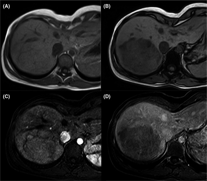
FIGURE 2.

H‐HCA in a 23‐year‐old woman. MR images show a large lesion in the right liver lobe. The MR shows the presence of diffuse fat deposition within the lesion (drop of the signal on opposed‐phase T1‐weighted image B—if compared to in phase image—A). The lesion slightly enhances on hepatic arterial phase (C) and shows washout on portal venous phase (D)
