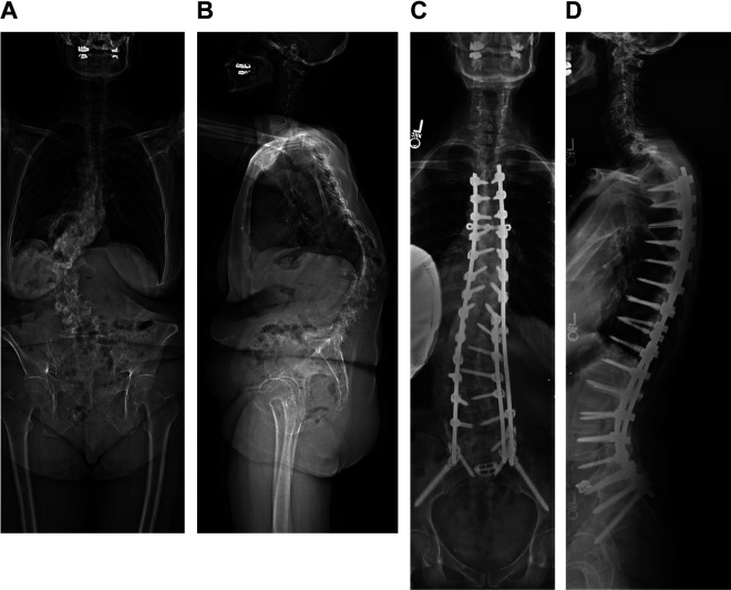
Figure 2.
67F presenting with several years of worsening back pain and deformity, refractory to conservative treatments. Preoperative bone density scans revealed osteoporosis; the patient underwent treatment with teriparatide in the months preceeding surgery. Preoperative standing radiographs (A, B), with baseline sagittal radiographic parameters included a sacral slope of 12°, pelvic tilt of 36°, pelvic incidence 47°, lumbar lordosis 16°, thoracolumbar junction (T10-L2) with 58° of kyphosis, and a C7 sagittal vertical axis (SVA) of 91 mm. The patient was instrumented from T3-pelvis, with Schwab grade 2 osteotomies from L1 to L4 and a transforaminal interbody fusion at L5/S1. The PLS was augmented at the top of the construct using the technique previously described. An increase in the proximal junctional angle from 2° of lordosis to 16° of kyphosis was first noted at the 6-week postoperative visit, which remained stable and asymptomatic through her 2-year postoperative radiographs (C, D).

