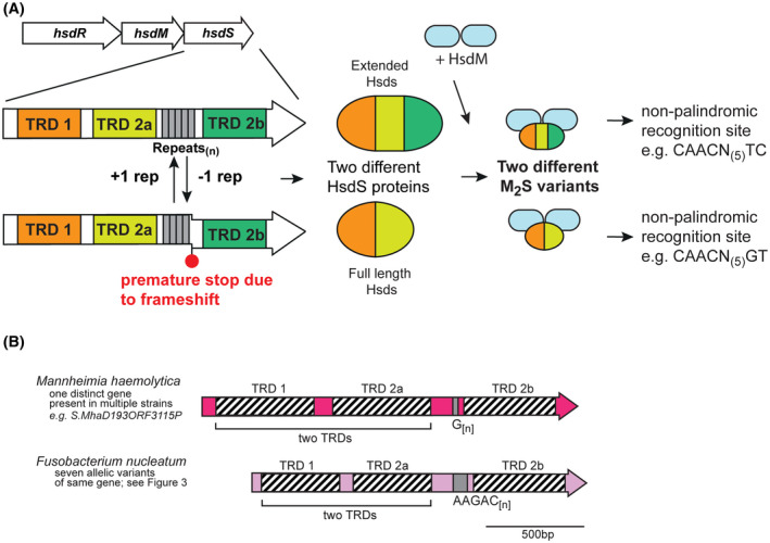
Figure 3.

A, Illustration of how phase‐variable switching of extended hsdS genes occurs. Extended hsdS genes contain three separate target recognition domains (TRDs; TRD 1 in orange at the 5′ end, a central TRD 2a in light green, and TRD 2b at the 3′ end in dark green). The SSR tract (gray boxes) is located between TRD 2a and TRD 2b. A frameshift mutation through loss or gain of repeat units in the SSR tract results in TRD 2b being out of frame with the rest of the gene, and expression of a full‐length HsdS protein consisting of TRD 1 + TRD 2a, analogous to that in Figure 1. However, if the SSR tract length results in read‐through to TRD 2b, a protein made up of all three TRDs (TRD 1 + 2a + 2b) is expressed. Following oligomerization with an HsdM dimer to form an active methyltransferase, the different HsdS protein subunits result in two different methyltransferase specificities. B, schematic representation of extended hsdS loci in Mannheimia haemolytica and Fusobacterium nucleatum. Colored arrows represent different genes, with color representing homology within each gene if more than one example of this gene is present in REBASE. Hatched boxes represent the locations of each TRD. The number of different hsdS genes is noted below each species. Unique examples are listed below each individual bacterial species where this hsdS gene is present
