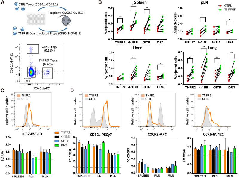
Figure 5.

TNFRSF costimulation increased Treg in vivo expansion and modified Treg tissue recirculation. Tregs, preactivated for 3 days with anti‐CD3/CD28 mAbs alone (control Tregs) or combined with TNFRSF costimulation (co‐stimulated Tregs), were cotransferred in equal numbers to assess their in vivo homeostasis 7 days later. (A) Experimental design (upper panel) and representative plot showing cotransferred Tregs identified with congenic markers (lower panel). (B) Proportions of coinjected Tregs in spleen, peripheral lymph nodes (pLN), liver, and lung. Each dot is a mouse and lines connect cells from the same mouse. Unpaired Mann–Whitney test was used. (C, D) Ki67, CD62L, CXCR3, and CCR6 expression among injected cells. Upper panels show representative histograms for control and TNFR2 costimulated Tregs in the spleen. Lower panels show the mean (±SD) of FC MFI expression of costimulated compared to control Tregs in spleen, pLN, and mesenteric (MLN) LN. Data were obtained from two independent experiments with six mice per group in total. From 100 to 500 cells were analyzed per gated Tregs.
