Figure 7.

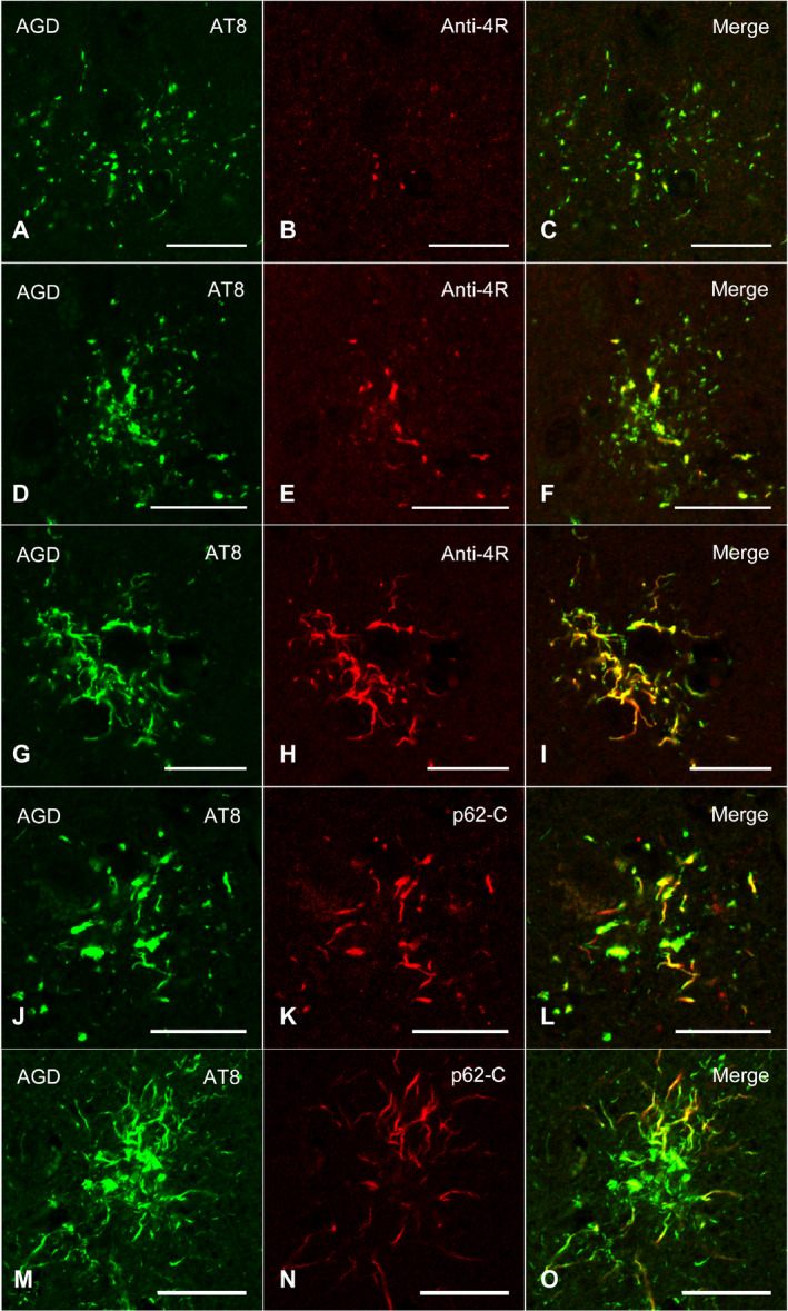
Confocal double‐immunofluorescence of the combination of AT8 (A, D, G) and anti‐4R tau antibodies (B, E, H), and the combination of AT8 (J, M) and p62‐C (K, N) in the frontal cortex in an AGD case. A–C, D–F. GFAs. An AT8 epitope is partially colocalized with an anti‐4R tau epitope. G–I. A TA. An anti‐4R tau epitope is often colocalized with an AT8 epitope. J–L. A GFA. Some GFAs having not only fine tau granules, but also coarse tau granules showed the colocalization of epitopes of AT8 and p62‐C. M–O. A TA. Some TAs also rarely showed the colocalization of epitopes of AT8 and p62‐C. C, F, I, L, O. Merged images. All scale bars = 20 μm.
